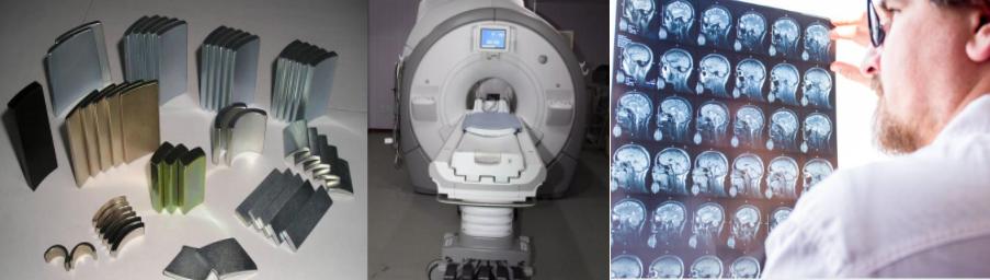
How Does Magnet Work In Magnetic Resonance CT?
The magnet is extremely widely used in our daily life that is closely associated with our life, such as speakers, communication, household applications, medical apparatus and instruments, and so on. Modern medicine uses the principle of magnetic resonance made magnetic resonance CT, which is a greatly convenient medical treatment, now, here will tell you how a magnet works in magnetic resonance CT.
Magnet Working Principle In Magnetic Resonance CT
In magnetic resonance CT, it makes use of the principle of nuclear magnetic resonance to diagnose abnormal tissues in the human body and judge diseases, which is the nuclear magnetic resonance imaging technology we are more familiar with, the basic principle is as follows: the nucleus has a positive charge and spin motion. Normally, the spin axes of nuclear nuclei are arranged irregularly, but when they are placed in an external magnetic field, the nuclear spin spatial orientation will transfer disorder to order. The magnetization vector of the spin system increases gradually from zero, and when the system reaches equilibrium, the magnetization intensity will reach a stable value. If the nuclear spin system is subjected to external action, such as a certain frequency of radio frequency excited nucleus can cause a resonance effect. After the radio frequency pulse stops, the nucleus of the spin system has been intensified and can not maintain this state, and return to the original arrangement in the magnetic field, as well as release weak energy at the same time, then become the radio signals, all of these signals will be detected and spatially resolved, at last, the picture of the distribution of nuclei in motion is obtained.
How Magnetic Resonance CT Imaging Works
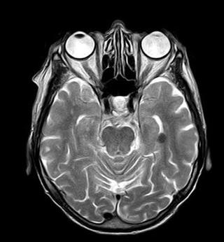
The characteristic of nuclear magnetic resonance is that flowing liquids do not generate signals, which is called the flow effect or the flow blank effect. Therefore, the blood vessels are grayish white tubular structures, while the blood is black without signals. This allows blood vessels to be easily separated from soft tissue. The normal medulla spinalis is surrounded by cerebrospinal fluid, which is black and has a white dura membrane set off by fat, making the spinal cord appear as a strong white signal structure. Magnetic resonance imaging has been applied to imaging diagnosis of various systems throughout the body. The best results are obtained from the brain, and spinal cord, heart blood vessels, joints bones, soft tissue, pelvis, and so on.
What’s more, for cardiovascular diseases, it is not only possible to observe the anatomical changes of various chambers, large blood vessels, and valves, but also to perform ventricular analysis for qualitative and semi quantitative diagnosis. Importantly, it provides multiple sectional views with high spatial resolution, and shows the overall picture of the heart and lesions, as well as their relationship with surrounding structures, which is superior to other X-ray imaging, two-dimensional ultrasound, radionuclide, and CT examinations.
Conclusion
This article tells you how magnets work and the working principle in Magnetic resonance CT, if this is helpful to you, and if you want to know more about the application of magnets in different fields and different use of different magnets, scan and read the product pages on this website where you can get an answer. Or if you are looking for a qualified magnet supplier to purchase a batch of magnets, KENENG is a good choice.
Related Products
No posts